Review


polyclonal rabbit anti spcs2  (Proteintech)


Bioz Verified Symbol Proteintech is a verified supplier  
  • Logo
  • About
  • News
  • Press Release
  • Team
  • Advisors
  • Partners
  • Contact
  • Bioz Stars
  • Bioz vStars
  • 93

    Structured Review

    Proteintech polyclonal rabbit anti spcs2
    Polyclonal Rabbit Anti Spcs2, supplied by Proteintech, used in various techniques. Bioz Stars score: 93/100, based on 6 PubMed citations. ZERO BIAS - scores, article reviews, protocol conditions and more
    https://www.bioz.com/product/anti+spcs2+rabbit+polyclonal+antibody/SPCS2+Antibody/pm36314723-407-117-121
    Average 93 stars, based on 6 article reviews
    polyclonal rabbit anti spcs2 - by Bioz Stars, 2026-09
    93/100 stars

    Images

    Related Articles

    Saline:

    Article Title: Cleavage of the Jaw1 C-terminal region enhances its augmentative effect on the Ca 2+ release via inositol 1,4,5-trisphosphate receptors
    Article Snippet: For the detection of SEC11A, SPCS1, and SPCS2, the samples were blotted onto a AmershamTM HybondTM P 0.2-μm PVDF membrane (#10600058; Cytiva, Marlborough, MA, USA). .. The membranes were blocked in 3% skim milk (#190-12865; FUJIFILM Wako Pure Chemical Corporation, Tokyo, Japan) diluted with Tris-buffered saline (TBS) (20 mM Tris–HCl pH 7.6 and 137 mM NaCl) containing 0.1% Tween-20 (TBS-T) for 1 h. After washing the membrane with TBS-T, it was reacted with the following primary antibodies diluted with 1% skim milk/TBS-T overnight at 4°C: anti-FLAG mouse antibody (1:1000) (#014-23383; FUJIFILM Wako Pure Chemical Corporation, Tokyo, Japan), anti-GAPDH mouse monoclonal antibody (1:1000) (#016-25523; FUJIFILM Wako Pure Chemical Corporation, Tokyo, Japan), anti-Jaw1 Coil rat antibody (1:500 or 1:1000) (produced in our laboratory as previously described ( Kozono et al ., 2018 )), anti-SEC11A rabbit polyclonal antibody (1:1000) (#14753-1-AP; Proteintech, Wuhan, Hubei, China), anti-SPCS1 rabbit polyclonal antibody (1:1000) (#11847-1-AP; Proteintech, Wuhan, Hubei, China), anti-SPCS2 rabbit polyclonal antibody (1:1000) (#14872-1-AP; Proteintech, Wuhan, Hubei, China), anti-Jaw1 N rabbit antibody (1:1000) (produced in our laboratory as described ( Okumura et al ., 2022 )), anti-IP 3 R1 rabbit antibody (1:1000) (#A7905; ABclonal, Wuhan, China), anti-IP 3 R2 mouse antibody (1:1000) (#sc-398434; Santa Cruz Biotechnology, CA, USA), and anti-IP 3 R3 mouse antibody (1:1000) (#610312;BD Bioscience, CA, USA). .. After washing the membrane with TBS-T, it was incubated with secondary antibodies diluted into 1:5000 with TBS-T for 1 h: Anti-Rat IgG, HRP-Linked Whole Ab Goat (#NA935; Cytiva, Marlborough, MA, USA), Anti-Rabbit IgG, HRP-Linked Whole Ab Donkey (#NA934; Cytiva, Marlborough, MA, USA), and anti-mouse IgG, HRP-Linked Whole Ab Sheep (#NA931; Cytiva, Marlborough, MA, USA).

    Membrane:

    Article Title: Cleavage of the Jaw1 C-terminal region enhances its augmentative effect on the Ca 2+ release via inositol 1,4,5-trisphosphate receptors
    Article Snippet: For the detection of SEC11A, SPCS1, and SPCS2, the samples were blotted onto a AmershamTM HybondTM P 0.2-μm PVDF membrane (#10600058; Cytiva, Marlborough, MA, USA). .. The membranes were blocked in 3% skim milk (#190-12865; FUJIFILM Wako Pure Chemical Corporation, Tokyo, Japan) diluted with Tris-buffered saline (TBS) (20 mM Tris–HCl pH 7.6 and 137 mM NaCl) containing 0.1% Tween-20 (TBS-T) for 1 h. After washing the membrane with TBS-T, it was reacted with the following primary antibodies diluted with 1% skim milk/TBS-T overnight at 4°C: anti-FLAG mouse antibody (1:1000) (#014-23383; FUJIFILM Wako Pure Chemical Corporation, Tokyo, Japan), anti-GAPDH mouse monoclonal antibody (1:1000) (#016-25523; FUJIFILM Wako Pure Chemical Corporation, Tokyo, Japan), anti-Jaw1 Coil rat antibody (1:500 or 1:1000) (produced in our laboratory as previously described ( Kozono et al ., 2018 )), anti-SEC11A rabbit polyclonal antibody (1:1000) (#14753-1-AP; Proteintech, Wuhan, Hubei, China), anti-SPCS1 rabbit polyclonal antibody (1:1000) (#11847-1-AP; Proteintech, Wuhan, Hubei, China), anti-SPCS2 rabbit polyclonal antibody (1:1000) (#14872-1-AP; Proteintech, Wuhan, Hubei, China), anti-Jaw1 N rabbit antibody (1:1000) (produced in our laboratory as described ( Okumura et al ., 2022 )), anti-IP 3 R1 rabbit antibody (1:1000) (#A7905; ABclonal, Wuhan, China), anti-IP 3 R2 mouse antibody (1:1000) (#sc-398434; Santa Cruz Biotechnology, CA, USA), and anti-IP 3 R3 mouse antibody (1:1000) (#610312;BD Bioscience, CA, USA). .. After washing the membrane with TBS-T, it was incubated with secondary antibodies diluted into 1:5000 with TBS-T for 1 h: Anti-Rat IgG, HRP-Linked Whole Ab Goat (#NA935; Cytiva, Marlborough, MA, USA), Anti-Rabbit IgG, HRP-Linked Whole Ab Donkey (#NA934; Cytiva, Marlborough, MA, USA), and anti-mouse IgG, HRP-Linked Whole Ab Sheep (#NA931; Cytiva, Marlborough, MA, USA).

    Produced:

    Article Title: Cleavage of the Jaw1 C-terminal region enhances its augmentative effect on the Ca 2+ release via inositol 1,4,5-trisphosphate receptors
    Article Snippet: For the detection of SEC11A, SPCS1, and SPCS2, the samples were blotted onto a AmershamTM HybondTM P 0.2-μm PVDF membrane (#10600058; Cytiva, Marlborough, MA, USA). .. The membranes were blocked in 3% skim milk (#190-12865; FUJIFILM Wako Pure Chemical Corporation, Tokyo, Japan) diluted with Tris-buffered saline (TBS) (20 mM Tris–HCl pH 7.6 and 137 mM NaCl) containing 0.1% Tween-20 (TBS-T) for 1 h. After washing the membrane with TBS-T, it was reacted with the following primary antibodies diluted with 1% skim milk/TBS-T overnight at 4°C: anti-FLAG mouse antibody (1:1000) (#014-23383; FUJIFILM Wako Pure Chemical Corporation, Tokyo, Japan), anti-GAPDH mouse monoclonal antibody (1:1000) (#016-25523; FUJIFILM Wako Pure Chemical Corporation, Tokyo, Japan), anti-Jaw1 Coil rat antibody (1:500 or 1:1000) (produced in our laboratory as previously described ( Kozono et al ., 2018 )), anti-SEC11A rabbit polyclonal antibody (1:1000) (#14753-1-AP; Proteintech, Wuhan, Hubei, China), anti-SPCS1 rabbit polyclonal antibody (1:1000) (#11847-1-AP; Proteintech, Wuhan, Hubei, China), anti-SPCS2 rabbit polyclonal antibody (1:1000) (#14872-1-AP; Proteintech, Wuhan, Hubei, China), anti-Jaw1 N rabbit antibody (1:1000) (produced in our laboratory as described ( Okumura et al ., 2022 )), anti-IP 3 R1 rabbit antibody (1:1000) (#A7905; ABclonal, Wuhan, China), anti-IP 3 R2 mouse antibody (1:1000) (#sc-398434; Santa Cruz Biotechnology, CA, USA), and anti-IP 3 R3 mouse antibody (1:1000) (#610312;BD Bioscience, CA, USA). .. After washing the membrane with TBS-T, it was incubated with secondary antibodies diluted into 1:5000 with TBS-T for 1 h: Anti-Rat IgG, HRP-Linked Whole Ab Goat (#NA935; Cytiva, Marlborough, MA, USA), Anti-Rabbit IgG, HRP-Linked Whole Ab Donkey (#NA934; Cytiva, Marlborough, MA, USA), and anti-mouse IgG, HRP-Linked Whole Ab Sheep (#NA931; Cytiva, Marlborough, MA, USA).


    Similar Products

    93
    Proteintech polyclonal rabbit anti spcs2
    Polyclonal Rabbit Anti Spcs2, supplied by Proteintech, used in various techniques. Bioz Stars score: 93/100, based on 1 PubMed citations. ZERO BIAS - scores, article reviews, protocol conditions and more
    https://www.bioz.com/product/anti+spcs2+rabbit+polyclonal+antibody/SPCS2+Antibody/pm36314723-407-117-121
    Average 93 stars, based on 1 article reviews
    polyclonal rabbit anti spcs2 - by Bioz Stars, 2026-09
    93/100 stars
      Buy from Supplier

    93
    Proteintech anti spcs2 rabbit polyclonal antibody
    Flp-In T-REx HEK293 Hu Jaw1 cells were treated with siRNA against spcs1, <t>spcs2</t> , and spcs3 for 48 h. After 24 h from the start of siRNA treatment, the cells were treated with Dox for 24 h. A-C ) Graphs showing the relative expression levels to NT of spcs1 (A), spcs2 (B), and spcs3 (C) mRNA to gapdh measured by RT-qPCR. D ) The cell lysates were subjected to western blotting. E-G ) Graphs showing the relative expression level of SPCS1 (E), SPCS2 (F), and SEC11A (G) in (D). H ) Graph showing the percentage of Jaw1 C-terminal cleavage in (D). A-C, E-H ) The averages of three independent experiments per condition are shown in the graphs. Error bar shows ±SD, “n.s.”, not significant; * P < 0.05; ** P < 0.01; *** P < 0.001; **** P < 0.0001. Statistics: one-way ANOVA followed by Dunnett’s multiple comparison test.
    Anti Spcs2 Rabbit Polyclonal Antibody, supplied by Proteintech, used in various techniques. Bioz Stars score: 93/100, based on 1 PubMed citations. ZERO BIAS - scores, article reviews, protocol conditions and more
    https://www.bioz.com/product/anti+spcs2+rabbit+polyclonal+antibody/SPCS2+Antibody/bio_rxiv__2022__12__10__519934-209-125-131
    Average 93 stars, based on 1 article reviews
    anti spcs2 rabbit polyclonal antibody - by Bioz Stars, 2026-09
    93/100 stars
      Buy from Supplier

    Image Search Results


    Flp-In T-REx HEK293 Hu Jaw1 cells were treated with siRNA against spcs1, spcs2 , and spcs3 for 48 h. After 24 h from the start of siRNA treatment, the cells were treated with Dox for 24 h. A-C ) Graphs showing the relative expression levels to NT of spcs1 (A), spcs2 (B), and spcs3 (C) mRNA to gapdh measured by RT-qPCR. D ) The cell lysates were subjected to western blotting. E-G ) Graphs showing the relative expression level of SPCS1 (E), SPCS2 (F), and SEC11A (G) in (D). H ) Graph showing the percentage of Jaw1 C-terminal cleavage in (D). A-C, E-H ) The averages of three independent experiments per condition are shown in the graphs. Error bar shows ±SD, “n.s.”, not significant; * P < 0.05; ** P < 0.01; *** P < 0.001; **** P < 0.0001. Statistics: one-way ANOVA followed by Dunnett’s multiple comparison test.

    Journal: bioRxiv

    Article Title: Cleavage of the Jaw1 C-terminal region enhances its augmentative effect on the Ca 2+ release via inositol 1,4,5-trisphosphate receptors

    doi: 10.1101/2022.12.10.519934

    Figure Lengend Snippet: Flp-In T-REx HEK293 Hu Jaw1 cells were treated with siRNA against spcs1, spcs2 , and spcs3 for 48 h. After 24 h from the start of siRNA treatment, the cells were treated with Dox for 24 h. A-C ) Graphs showing the relative expression levels to NT of spcs1 (A), spcs2 (B), and spcs3 (C) mRNA to gapdh measured by RT-qPCR. D ) The cell lysates were subjected to western blotting. E-G ) Graphs showing the relative expression level of SPCS1 (E), SPCS2 (F), and SEC11A (G) in (D). H ) Graph showing the percentage of Jaw1 C-terminal cleavage in (D). A-C, E-H ) The averages of three independent experiments per condition are shown in the graphs. Error bar shows ±SD, “n.s.”, not significant; * P < 0.05; ** P < 0.01; *** P < 0.001; **** P < 0.0001. Statistics: one-way ANOVA followed by Dunnett’s multiple comparison test.

    Article Snippet: The membranes were blocked in 3% skim milk (#190-12865; FUJIFILM Wako Pure Chemical Corporation, Tokyo, Japan) diluted with Tris-buffered saline (TBS) (20 mM Tris–HCl pH 7.6 and 137 mM NaCl) containing 0.1% Tween-20 (TBS-T) for 1 h. After washing the membrane with TBS-T, it was reacted with the following primary antibodies diluted with 1% skim milk/TBS-T overnight at 4°C: anti-FLAG mouse antibody (1:1000) (#014-23383; FUJIFILM Wako Pure Chemical Corporation, Tokyo, Japan), anti-GAPDH mouse monoclonal antibody (1:1000) (#016-25523; FUJIFILM Wako Pure Chemical Corporation, Tokyo, Japan), anti-Jaw1 Coil rat antibody (1:500 or 1:1000) (produced in our laboratory as previously described ( Kozono et al ., 2018 )), anti-SEC11A rabbit polyclonal antibody (1:1000) (#14753-1-AP; Proteintech, Wuhan, Hubei, China), anti-SPCS1 rabbit polyclonal antibody (1:1000) (#11847-1-AP; Proteintech, Wuhan, Hubei, China), anti-SPCS2 rabbit polyclonal antibody (1:1000) (#14872-1-AP; Proteintech, Wuhan, Hubei, China), anti-Jaw1 N rabbit antibody (1:1000) (produced in our laboratory as described ( Okumura et al ., 2022 )), anti-IP 3 R1 rabbit antibody (1:1000) (#A7905; ABclonal, Wuhan, China), anti-IP 3 R2 mouse antibody (1:1000) (#sc-398434; Santa Cruz Biotechnology, CA, USA), and anti-IP 3 R3 mouse antibody (1:1000) (#610312;BD Bioscience, CA, USA).

    Techniques: Expressing, Quantitative RT-PCR, Western Blot, Comparison

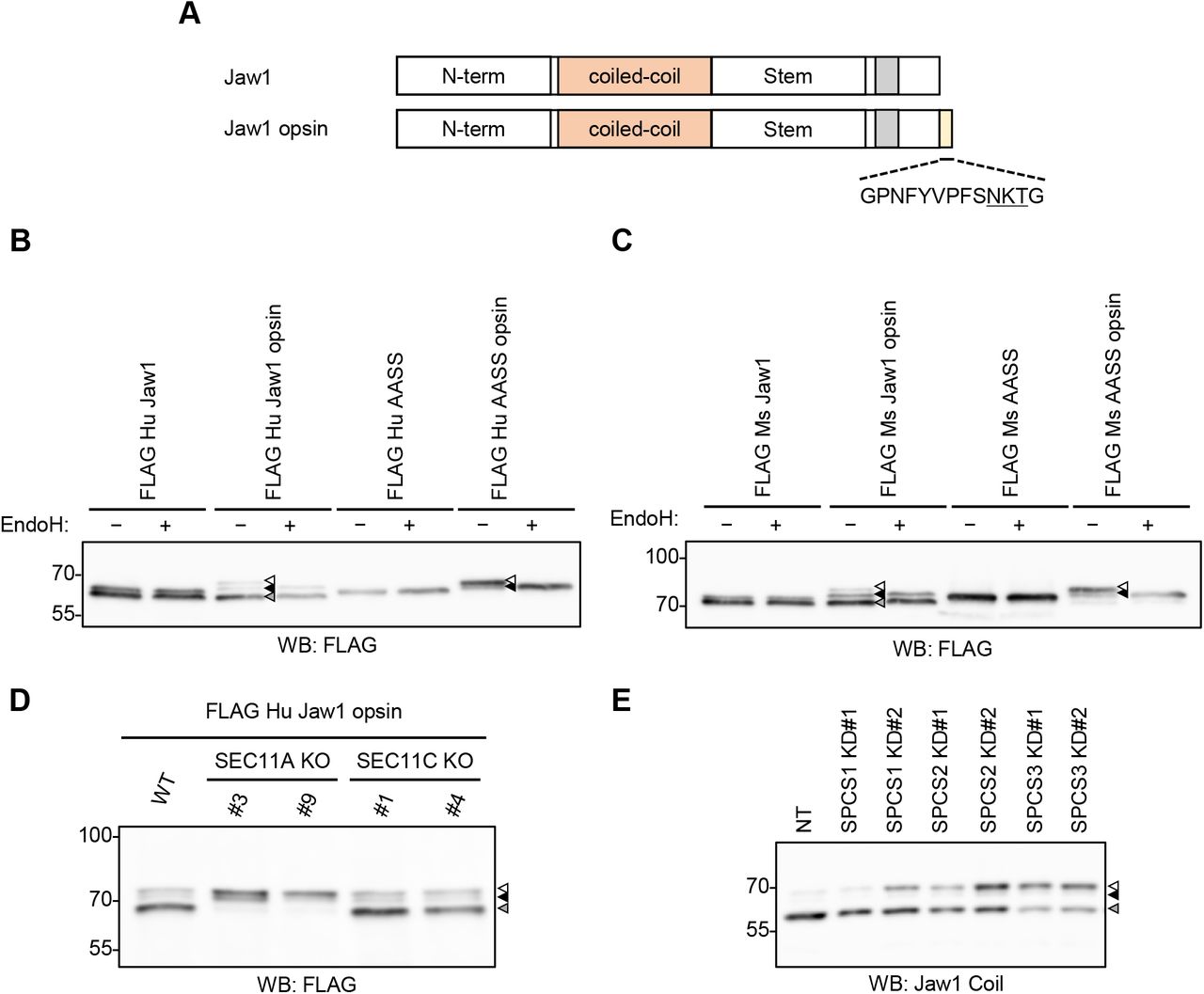
    A ) Schematic representation of Jaw1 and Jaw1 opsin. The opsin tag was added to its C-terminal end. B, C ) FLAG Hu Jaw1, FLAG Hu Jaw1 opsin, FLAG Hu AASS, and FLAG Hu AASS opsin (B) and FLAG Ms Jaw1, FLAG Ms Jaw1 opsin, FLAG Ms AASS, and FLAG Ms AASS opsin (C) were expressed in HEK293 cells by transfection. After incubation for 24 h, the lysates samples were treated with or without EndoH and subjected to western blotting using an anti-FLAG mouse antibody. D ) FLAG Hu Jaw1 opsin was expressed in SEC11A KO #3 and #9 and SEC11C KO #1 and #4 cells by transfection. After incubation for 24 h, the lysates were subjected to western blotting using an anti-FLAG mouse antibody. E ) Flp-In T-REx HEK293 Hu Jaw1 opsin cells were treated with siRNA against spcs1, spcs2 , and spcs3 for 48 h. After 24 h from the start of siRNA treatment, the cells were treated with Dox for 24 h. After that, the lysates were subjected to western blotting using an anti-Jaw1 Coil antibody. Hu Jaw1 opsin expressed by the treatment with Dox in this cell does not bear the N-terminal tags unlike FLAG Hu Jaw1 opsin in (B) and (D), thereby, an anti-Jaw1 Coil antibody but not an anti-FLAG mouse antibody was used. B-E ) Open triangles, the bands of ER-inserted uncleaved Jaw1 with N -linked glycosylation; closed triangles, the bands of the pre-inserted Jaw1(black) and cleaved Jaw1 (gray). The representative blot images from three independent experiments with similar results are shown.

    Journal: bioRxiv

    Article Title: Cleavage of the Jaw1 C-terminal region enhances its augmentative effect on the Ca 2+ release via inositol 1,4,5-trisphosphate receptors

    doi: 10.1101/2022.12.10.519934

    Figure Lengend Snippet: A ) Schematic representation of Jaw1 and Jaw1 opsin. The opsin tag was added to its C-terminal end. B, C ) FLAG Hu Jaw1, FLAG Hu Jaw1 opsin, FLAG Hu AASS, and FLAG Hu AASS opsin (B) and FLAG Ms Jaw1, FLAG Ms Jaw1 opsin, FLAG Ms AASS, and FLAG Ms AASS opsin (C) were expressed in HEK293 cells by transfection. After incubation for 24 h, the lysates samples were treated with or without EndoH and subjected to western blotting using an anti-FLAG mouse antibody. D ) FLAG Hu Jaw1 opsin was expressed in SEC11A KO #3 and #9 and SEC11C KO #1 and #4 cells by transfection. After incubation for 24 h, the lysates were subjected to western blotting using an anti-FLAG mouse antibody. E ) Flp-In T-REx HEK293 Hu Jaw1 opsin cells were treated with siRNA against spcs1, spcs2 , and spcs3 for 48 h. After 24 h from the start of siRNA treatment, the cells were treated with Dox for 24 h. After that, the lysates were subjected to western blotting using an anti-Jaw1 Coil antibody. Hu Jaw1 opsin expressed by the treatment with Dox in this cell does not bear the N-terminal tags unlike FLAG Hu Jaw1 opsin in (B) and (D), thereby, an anti-Jaw1 Coil antibody but not an anti-FLAG mouse antibody was used. B-E ) Open triangles, the bands of ER-inserted uncleaved Jaw1 with N -linked glycosylation; closed triangles, the bands of the pre-inserted Jaw1(black) and cleaved Jaw1 (gray). The representative blot images from three independent experiments with similar results are shown.

    Article Snippet: The membranes were blocked in 3% skim milk (#190-12865; FUJIFILM Wako Pure Chemical Corporation, Tokyo, Japan) diluted with Tris-buffered saline (TBS) (20 mM Tris–HCl pH 7.6 and 137 mM NaCl) containing 0.1% Tween-20 (TBS-T) for 1 h. After washing the membrane with TBS-T, it was reacted with the following primary antibodies diluted with 1% skim milk/TBS-T overnight at 4°C: anti-FLAG mouse antibody (1:1000) (#014-23383; FUJIFILM Wako Pure Chemical Corporation, Tokyo, Japan), anti-GAPDH mouse monoclonal antibody (1:1000) (#016-25523; FUJIFILM Wako Pure Chemical Corporation, Tokyo, Japan), anti-Jaw1 Coil rat antibody (1:500 or 1:1000) (produced in our laboratory as previously described ( Kozono et al ., 2018 )), anti-SEC11A rabbit polyclonal antibody (1:1000) (#14753-1-AP; Proteintech, Wuhan, Hubei, China), anti-SPCS1 rabbit polyclonal antibody (1:1000) (#11847-1-AP; Proteintech, Wuhan, Hubei, China), anti-SPCS2 rabbit polyclonal antibody (1:1000) (#14872-1-AP; Proteintech, Wuhan, Hubei, China), anti-Jaw1 N rabbit antibody (1:1000) (produced in our laboratory as described ( Okumura et al ., 2022 )), anti-IP 3 R1 rabbit antibody (1:1000) (#A7905; ABclonal, Wuhan, China), anti-IP 3 R2 mouse antibody (1:1000) (#sc-398434; Santa Cruz Biotechnology, CA, USA), and anti-IP 3 R3 mouse antibody (1:1000) (#610312;BD Bioscience, CA, USA).

    Techniques: Transfection, Incubation, Western Blot, Glycoproteomics